Error: No layouts found
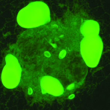
The ability to visualize proteins and other molecules using green fluorescent protein (GFP) and other FP “tags” has revolutionized light microscopy. But many FPs can weakly bind other copies of themselves, thereby interfering with experiments. Here, GFP was attached to a membrane protein in the endoplasmic reticulum (ER) of a cell. The GFP interactions have distorted the ER’s spaghetti-like tubules, causing them to expand, stack and form large bright structures. Erik L. Snapp, Ph.D., associate professor of anatomy & structural biology, and graduate student Lindsey Costantini have developed a visual assay to determine whether an FP has unwanted binding properties that can cause inappropriate interactions—and ruin someone’s experimental results.
Image credit: Erik L. Snapp, Ph.D.