 Image courtesy of David Entenberg, Ph.D., co-corresponding author of the Nature Reviews Cancer paper, associate professor of pathology, and co-director of the Gruss Lipper Biophotonics Center.
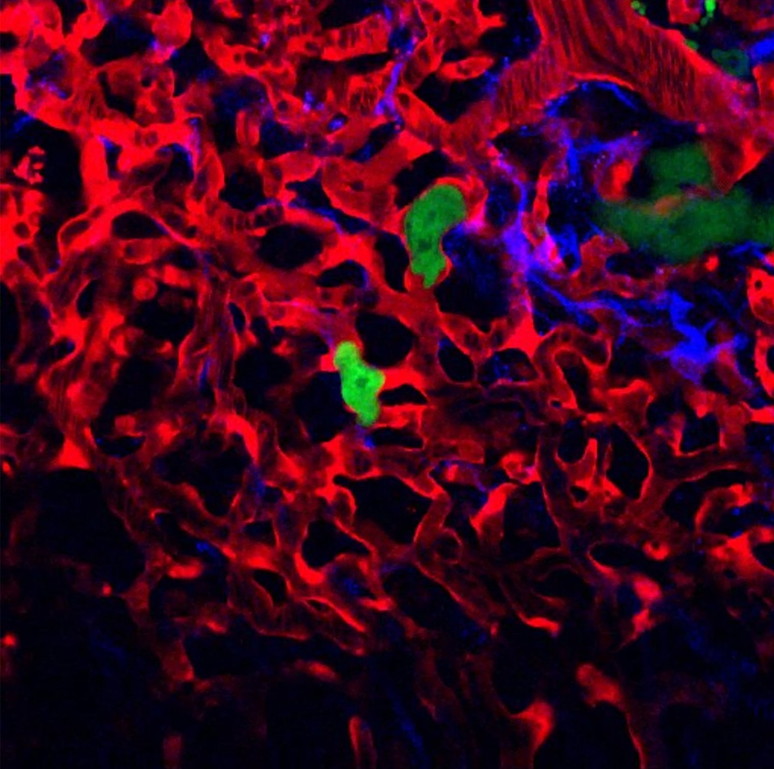
Some two-thirds of cancer deaths result from metastasis—when cells break away from a primary cancer and travel via the bloodstream to form new tumors in other parts of the body. Researchers in Einstein’s Gruss Lipper Biophotonics Center have pioneered the use of intravital imaging (IVI) to better understand how metastasis progresses and to develop novel therapies to halt it. As the researchers describe in the January 2023 issue of Nature Reviews Cancer, IVI can generate real-time subcellular-resolution images of single slices of intact tissues in live animals for several weeks; combining IVI with multiphoton microscopy and an implanted window enables internal organs to be viewed. At the center of this image of a live mouse lung are two tumor cells (green) from a breast tumor that have metastasized to the vessels (red) of the mouse’s lung. To their right is a cluster of growing tumor cells (also green) that have extravasated (exited the vasculature and entered lung tissue). Small green dots at the extreme top right are fragments of dead or dying tumor cells. Blue areas in the image are collagen fibers.