Hillary Guzik grew up in Syracuse, NY, loving photography and science the reason her early photos tended to exclude friends and family and instead featured insects, strands of human hair and even playground jungle gyms. “I’ve always liked things with repetitive patterns in them,” she explains.
A research technician in Einstein’s Analytical Imaging Facility (AIF), Ms. Guzik earned a B.S. degree in biomedical photographic communications from the Rochester Institute of Technology. “When I found that major at RIT,” she says, “it was like, ‘That’s what I’m going to do for the rest of my life!’” She learned theoretical and practical microscopy applications—everything from sample preparation to imaging acquisition to creating scientific posters. “So when I came here in 2009,” she adds, “I had many of the skills I needed.”
One of Ms. Guzik’s tasks in the AIF involves teaching people at Einstein how to use light microscopes, including epi-fluorescence, super-resolution, brightfield and confocal models—the latter a specialized type of light microscope that uses lasers to scan across specimens to form images. She and her AIF colleagues also offer image analysis—for example, quantifying fluorescent intensities to see whether certain tumor-cell proteins are over- or underexpressed.
“Most of the work in the AIF is medically oriented,” explains Ms. Guzik. “Researchers might use the facility to compare breast cancer treatments by viewing tumor cells that have been exposed to different therapies. Or researchers studying multiple sclerosis might assess various drugs to see if they help grow or regrow axons in nerves.”
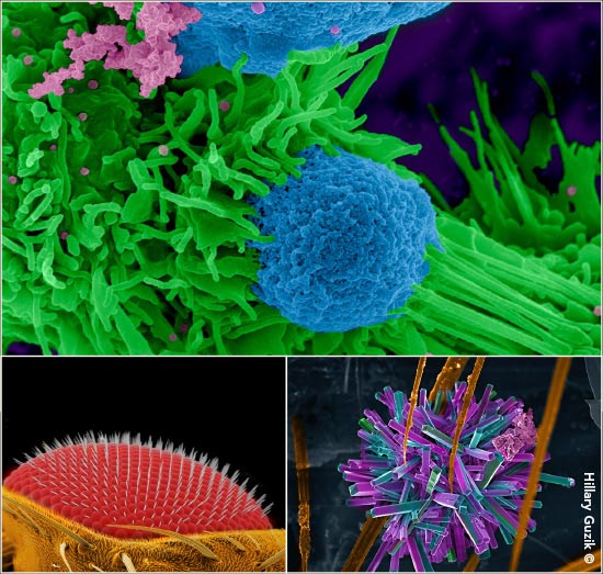
Ms. Guzik particularly likes applying her Photoshop skills to color scanning and transmission electron microscope images, both those she takes herself and those on which she collaborates. In 2014, her work colorizing originally black-and-white images garnered kudos when she partnered with postdoctoral fellow Sabriya Stukes. They won first prize in the annual BioArt photography contest held by the Federation of American Societies for Experimental Biology to showcase “the beauty and excitement of biological research.” Their vibrant image (at right) depicts a tug-of-war between a fungal cell and two macrophages in a mouse.
“When I’m coloring these images, there are not many rules,” she says. “A cell’s nucleus is generally colored blue, but I basically use whatever colors I like!”
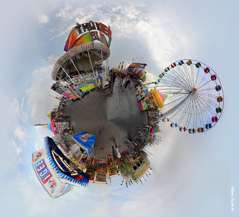
When not at work, Ms. Guzik likes being outdoors, hiking or rock climbing with her husband, Paul Jung—and still thinking in pictures. She especially enjoys creating 360-degree panoramic views of landscapes, towns and other scenes that she calls “small worlds.” Rather than lugging around a tripod, she simply takes her Nikon digital camera to a location and, standing in a central spot, turns slowly while taking 20 to 40 photos, being careful to overlap the images. She then uses Photoshop to “stitch” the images together and connect the left and right ends. “Technically,” she says, “it’s pretty easy to do, but finding the perfect spot is tough.”
Harking back to her early interest in insects, Ms. Guzik has a master’s degree in entomology from the University of Nebraska. Many insects are vectors for disease, and she’d like to study mosquitoes that transmit viral diseases such as West Nile, Zika and yellow fever.
 “Small world” photo of a state fair amusement park.
 Clockwise, above: Award-winning photo of macrophages; debris on a termite setae; up-close view of a Drosophila eye.
[ngg_images source=”galleries” container_ids=”2″ display_type=”photocrati-nextgen_basic_thumbnails” override_thumbnail_settings=”1″ thumbnail_width=”160″ thumbnail_height=”100″ thumbnail_crop=”1″ images_per_page=”20″ number_of_columns=”0″ ajax_pagination=”0″ show_all_in_lightbox=”0″ use_imagebrowser_effect=”0″ show_slideshow_link=”0″ slideshow_link_text=”[Show slideshow]” template=”/Users/aetherworks/Local Sites/aecm-magazine-migration/app/public-staging/wp-content/plugins/nextgen-gallery/products/photocrati_nextgen/modules/ngglegacy/view/gallery-caption.php” order_by=”sortorder” order_direction=”ASC” returns=”included” maximum_entity_count=”500″]