 Visualizing the bile canaliculi, the tiny channels into which hepatocytes (the predominant cell type in the liver) secrete bile.
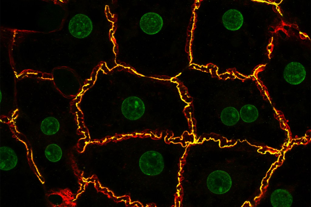
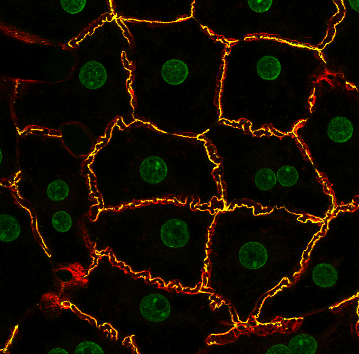
Hepatocytes—the predominant cell type in the liver—are polarized epithelial cells: Their basolateral domains face blood vessels and absorb substances from the blood, while their apical domains form the linings of the bile canalicular network—the tiny channels into which hepatocytes secrete bile, a detergent-like substance that breaks down fats in the small intestine.
Francisco Lazaro-Dieguez, Ph.D., staff scientist in the laboratory of Anne Muesch, Ph.D., studies the molecular mechanisms involved in maintaining the bile canaliculi; the maintenance of these channels is compromised in important liver diseases including non-alcoholic fatty liver disease and hepatitis. Dr. Lazaro-Dieguez made this image, which shows isolated rat hepatocytes manipulated to form canalicular networks similar to those in vivo. Hepatocyte nuclei are shown in green. To visualize the bile canaliculi, the tight-junction protein zonula occludens-1 (yellow) and actin filaments (red) were stained. The multiprotein tight junctions help keep hepatocytes polarized by forming barriers separating their apical and basolateral domains. The image was one of the winners of the photo contest sponsored by ibidi, a biotechnology company, and is the March “Image of the Month” in the 2024 ibidi calendar. Dr. Muesch is a professor of developmental & molecular biology at Einstein.