Cell movement is essential for crucial biological processes. It can help heal wounds but also lead to the spread of cancer cells (metastasis). It’s triggered when actin protein molecules combine, or polymerize, into actin filaments. Growing actin filaments push the cell membrane outward, forming projections called lamellipodia that propel the cell.
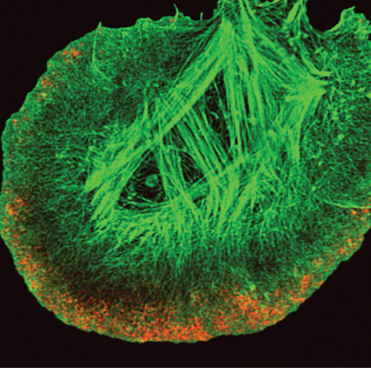
Visualizing this highly dynamic activity has proved challenging. To find the stains and preservation techniques that best capture actin polymerization within lamellipodia, researchers in Einstein’s Analytic Imaging Facility and the lab of John Condeelis, Ph.D., took motile cells from a rat breast cancer cell line, preserved (fixed) them in five different ways, and labeled the actin with nine different stains. The double-stained image shown here, using structural illumination microscopy, was judged the most accurate and detailed.
Phalloidin Alexa-488 (green) revealed most of the cell’s actin filament network. Anti-actin antibody AC15 Alexa-555 (red) showed actin staining that was limited to areas closer to the membrane.
Image credit: Vera DesMarais, Ph.D., and Robert Eddy, Ph.D.